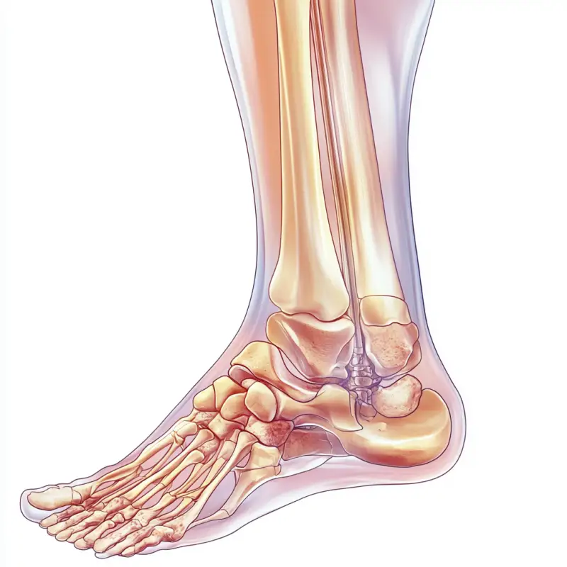
踝关节骨关节炎分期:1. 早期阶段: 症
提示词:Ankle osteoarthritis staging: 1 Early stage: Symptoms: Mild pain and stiffness, especially after physical activity. Imaging features: Joint space may be normal, with slight osteophyte formation. Image description: The joint gap is close to normal, displayed as a small gap line. Tiny osteophyte (small bone spurs or protrusions) begin to appear at the edge of the bone, but it is not severe. The structure of cartilage is basically intact, but there may be slight local irregularities. 2. Mid stage: Symptoms: Increased pain, limited mobility, and possible swelling. Imaging features: Narrowing of joint space, obvious osteophyte, and possible cartilage damage. Image description: The joint gap has significantly narrowed, with some areas even completely disappearing. The osteophyte has significantly increased in size and extended towards the joint, possibly protruding outward. The damage to cartilage begins to manifest, manifested as uneven or partially absent cartilage surface. 3. Late stage: Symptoms: persistent severe pain, severely limited joint function, and possible deformities. Imaging features: significant narrowing of joint space, osteophyte hyperplasia, possible osteoporosis or cysts on the joint surface. Image description: The joint gap almost disappears or is completely closed, displayed as joint surface contact. The osteophyte is large and hard, seriously affecting joint movement. There may be obvious osteoporosis, and there may be signs of cysts or complete loss of cartilage on the joint surface. Joint deformities may occur, such as changes in ankle joint shape or joint misalignment. Draw a schematic diagram of different stages of ankle arthritis based on the above information --v 6.1 --ar 1:1
素材来源:Midjourney官网
Copyright©2017 Midjourney9.com All Right
Reserved 版权所有:成都金翼云科技有限公司 蜀ICP备2023008999号